

 Part Two: Why Bones Become Fragile, Why Fractures Can Be Devastating, What Conventional Treatment May Be Missing, and Why There Is Hope
By Dr. Greg Fors, Chief Science Officer, Biospec Nutritionals, Clinic Director, Pain and Brain Healing Center
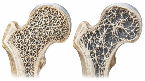
- Osteopenia and osteoporosis involve changes to both the amount of bone and its internal structure.
- Vertebral and hip fractures can affect posture, mobility, independence, and overall health.
- Bone is continually renewed: cells remove old bone while other cells build new bone.
- Inflammation, insulin resistance, and declining estrogen are among the factors that can influence bone health.
- CTX and P1NP can offer information about bone remodeling alongside DEXA scans.
Twenty years ago, my wife Carolyn received some disturbing news. She was only 54 years old, yet her first DEXA scan revealed bone density comparable to that of a 90-year-old woman, according to her primary care physician.
Think about that for a moment. A relatively young, otherwise healthy woman was facing a condition that could eventually rob her of her mobility, independence, and quality of life.
And there was another reason this diagnosis frightened us. Carolyn's grandmother had developed severe kyphosis from vertebral deterioration. I can still remember seeing her when she was approximately 65 years old, bent forward from the collapse of her spine. It was a painful reminder of what advanced osteoporosis can do to a person's body. Now Carolyn was facing a similar possibility.
Her GP recommended osteoporosis medications. But after Carolyn investigated the available drug treatments, their adverse effects, and their long-term implications, she made a decision.
She was not going to take those drugs.
I supported her decision, and I began exploring a more comprehensive approach to restoring and maintaining healthy bone.
Today, Carolyn is 74 years old. She has never experienced a spontaneous vertebral compression fracture, disabling kyphosis, or the progressive loss of independence we feared. Even more encouraging, her serial DEXA scans, performed approximately every two years, have documented improvements in bone density.
And she has accomplished this without ever taking conventional prescription osteoporosis medications. Instead, we have spent more than two decades addressing the nutritional, hormonal, metabolic, inflammatory, and lifestyle factors that influence bone health.
In the past few years, her advanced blood markers for osteoporosis have improved, providing another encouraging perspective. Her CTX, a marker of bone breakdown, has moved into the lower-normal range, while her P1NP, a marker of new bone collagen formation, has moved into the upper-normal range.
These results suggest a favorable pattern of bone remodeling and improved bone health, particularly when considered alongside her documented improvements on DEXA scans. I share Carolyn's experience because I believe we need to change the conversation about osteoporosis.
Too often, women are told that osteoporosis is simply an inevitable consequence of aging. They are offered medications with potentially serious adverse effects, yet receive relatively little guidance about the underlying biological processes responsible for their bone loss.
I believe women deserve a more comprehensive approach. And that begins with understanding what osteoporosis really is.
Osteopenia and Osteoporosis: What's Actually Happening to the Bones?
Most clinicians are familiar with the terms osteopenia and osteoporosis. But I think we sometimes become so focused on the DEXA numbers that we forget what is actually happening inside the skeleton.
Osteopenia means bone mineral density has declined below the expected range for healthy young adults. Osteoporosis represents more advanced deterioration of bone strength, involving reduced bone mass and disruption of the internal architecture of the skeleton.
A DEXA scan measures bone mineral density, usually at the lumbar spine and hip. The results are commonly expressed as a T-score. A T-score of −1.0 or higher is generally considered normal. Between −1.0 and −2.5 indicates osteopenia, while −2.5 or lower indicates osteoporosis in the populations for which these thresholds apply [1]. But here's the problem.
A DEXA scan tells us how much mineralized bone is present. It doesn't tell us everything about how strong that bone really is.
Healthy bone is a remarkable engineering structure. It contains a framework of collagen fibers reinforced with mineral crystals. The collagen provides toughness and flexibility, while the minerals provide stiffness and resistance to compression.
Think of reinforced concrete. The concrete provides rigidity, while the steel reinforcement helps prevent the structure from cracking apart under stress. Bone works somewhat similarly.
As osteoporosis progresses, the internal supporting structures become thinner. Some connections disappear altogether. The result is a skeleton that may look relatively normal from the outside but is becoming dangerously fragile on the inside. And the frightening part?
The first symptom may be a fracture.
When the Spine Begins to Collapse
Osteoporosis is often called the silent disease because it usually develops without pain or obvious symptoms. But once fractures begin, the consequences can be devastating.
Consider vertebral compression fractures. The vertebrae are the individual bones that form our spinal column. In advanced osteoporosis, these bones may become so fragile that ordinary daily activities can cause them to partially collapse.
A woman bends forward to pick something up. She lifts a grocery bag. She coughs. And suddenly she experiences severe back pain.
Sometimes the fracture is less dramatic. The front of the vertebral body gradually compresses without producing much initial pain. Over time, several vertebrae may collapse. The person begins losing height. The upper back becomes increasingly rounded because compression of the front of the vertebral body causes the vertebrae to become wedge-shaped. Eventually, the familiar stooped posture of permanent kyphosis develops.
But this is much more than a cosmetic problem. As the spine bends forward, the body's center of gravity changes. The back muscles must work harder simply to keep the person upright. Chronic muscular fatigue and pain develop.
Balance becomes more difficult. Walking becomes less stable. The risk of falling increases. In severe cases, spinal deformity can even interfere with normal chest expansion and breathing [2].
One vertebral fracture can start a cascade of additional fractures, chronic pain, disability, and loss of independence.
This is what Carolyn and I feared when she received her diagnosis. We had already seen what severe kyphosis had done to her grandmother. And we were determined to do everything possible to prevent that outcome.

Hip Fractures: When a Broken Bone Becomes a Life-Threatening Event
If vertebral fractures are frightening, hip fractures can be catastrophic. For an elderly person, a hip fracture often means hospitalization, surgery, prolonged rehabilitation, and a major loss of independence.
Many patients never regain their previous level of mobility. Some require walkers or assistance with everyday activities. Others can no longer live independently.
And the complications can be serious: blood clots, pneumonia, infections, delirium, muscle wasting, and cardiovascular events. Research has consistently demonstrated substantial mortality following hip fractures in older adults. Depending on the population studied, approximately 20–30% may die within the following year [3].
Not every death is directly caused by the fracture. But the fracture can begin a cascade of complications in an already vulnerable person.

Preventing a hip fracture may mean preserving not only someone's independence but also their life.
That's why I take osteoporosis so seriously. And it's why I believe we should be addressing the underlying biology long before the first fracture occurs.
Why Are So Many Women Frightened of Osteoporosis Medications?
Throughout my clinical career, I have encountered countless women who come into my office with osteopenia or osteoporosis. They are frightened about their bones. But they're also frightened about the medications they've been offered.
Some have experienced adverse effects themselves. Others have watched friends or family members struggle with complications. Many have investigated the drugs and simply don't want to take them. I understand their concerns.
Carolyn was one of those women. And considering the severity of her osteoporosis at age 54, she was certainly someone with a severely increased fracture risk.
But after reviewing the available treatments, she decided that the potential consequences were unacceptable to her.
I believe women deserve to understand exactly what these medications do, not just what they may accomplish on a DEXA scan, but what happens when we interfere with the normal biology of bone remodeling.
Bisphosphonates: Are We Suppressing a Process the Body Actually Needs?
Let's start with bisphosphonates, including alendronate (Fosamax) and zoledronic acid (Reclast). These medications work primarily by suppressing osteoclasts, the cells responsible for breaking down old bone.
And yes, at times, suppressing bone resorption can increase bone mineral density. But here's the question I always ask.
Why does the body have osteoclasts in the first place?
Are they simply troublemakers destroying our skeleton? Of course not! Osteoclasts perform an essential biological function. They remove old and damaged bone so that osteoblasts can replace it with healthy new bone.
That's how the skeleton maintains itself. When we profoundly suppress that process over long periods, we also interfere with the body's ability to repair accumulated microscopic damage.
This is one proposed mechanism behind atypical femoral fractures, an uncommon but serious complication associated with prolonged antiresorptive therapy [4].
These fractures are particularly disturbing because they may occur in the shaft of the thighbone with little or no trauma. Some patients experience persistent thigh or groin pain before the fracture occurs. Think about the irony.
A medication prescribed by your doctor to prevent fractures can contribute to an entirely different type of fracture. And that's not the only bone concern. Bisphosphonates have also been associated with osteonecrosis of the jaw, a potentially serious condition in which an area of jawbone fails to heal normally [5].
Furthermore, oral formulations can cause reflux, esophageal irritation, abdominal discomfort, and occasionally severe musculoskeletal pain. Intravenous formulations may cause flu-like reactions, and kidney function must be considered.
Because the risks and benefits change over time, clinicians commonly reassess oral bisphosphonate therapy after approximately five years and intravenous therapy after approximately three years. Some patients are then taken off the medication for what is euphemistically called a "drug holiday" [1].
That alone should remind us that these are not biologically insignificant interventions.
Prolia: What Happens When the Injections Stop?
Now consider denosumab, marketed as Prolia. This medication blocks RANKL, a signaling protein required for the development and activity of osteoclasts.
By suppressing osteoclast activity, denosumab can produce substantial reductions in bone resorption and increases in bone mineral density. But what happens when the medication wears off? This is where things become particularly concerning. Bone resorption can rebound rapidly.
CTX levels may rise substantially above pretreatment values. Bone density can decline quickly, and some patients experience multiple vertebral compression fractures! [6]
Imagine taking a medication to prevent spinal fractures, only to discover that stopping it without another carefully planned treatment can expose you to accelerated bone loss and serious fracture risk.
That is not a minor consideration. Patients beginning denosumab need to understand that injections must remain on schedule and that discontinuation generally requires a carefully coordinated transition to another antiresorptive medication.
For women who are already reluctant to commit to long-term drug therapy, I can certainly understand why this creates concern.
Evenity: What About the Cardiovascular Warning?
Romosozumab, marketed as Evenity, takes a different approach. It inhibits sclerostin, a protein that normally restrains bone formation. This allows the medication to stimulate osteoblast activity while also reducing bone resorption.
An interesting mechanism. But Evenity carries a boxed warning concerning the potential risk of myocardial infarction, stroke, and cardiovascular death. That's not exactly reassuring to a woman who is already worried about her health.
The medication should not be initiated in patients who have experienced a heart attack or stroke within the preceding year. Furthermore, cardiovascular risk must be carefully evaluated before starting the medication [7].
Treatment is limited to 12 monthly doses under the approved regimen. Afterward, another osteoporosis medication is generally required to preserve the gains made.
Other osteoporosis medications have their own limitations. Teriparatide and abaloparatide stimulate bone formation but require injections and may cause dizziness, nausea, and disturbances in calcium metabolism. Raloxifene can increase the risk of venous thromboembolism.
The point is straightforward.
These medications are powerful interventions, and some of their adverse effects are serious. Women deserve to understand those risks before making their decisions.
Are We Building Better Bone, or Just Improving the DEXA Number?
Here's something I think deserves much more attention. Bone density is not the same thing as bone quality. A healthy skeleton needs adequate mineral content, strong collagen, sound microscopic architecture, and the ability to repair itself.
Antiresorptive medications can increase bone mineral density partly by reducing the amount of bone undergoing active remodeling and allowing existing bone to become more fully mineralized. But prolonged suppression of remodeling may also permit microscopic damage to accumulate in some circumstances [4,8]. So I believe we need to ask a bigger question.
Are we improving the health of the entire skeletal system, or are we becoming too focused on the DEXA number? Are we addressing the underlying causes of osteopenia and osteoporosis?
What about chronic inflammation?
What about insulin resistance?
What about declining estrogen and testosterone?
What about muscle mass, balance, protein intake, and nutritional status?
And what about the biological processes that determine whether old bone is being replaced by healthy new bone?
Shouldn't we be investigating those factors too? This is where I believe functional medicine has something important to contribute.
Bone Is Living Tissue: Meet the Demolition Crew and the Construction Crew
I often explain bone remodeling to patients by comparing it to a construction project. Imagine that your skeleton is a building that must remain strong for 80, 90, or even 100 years. You can't simply construct the building once and walk away.
Over time, parts of the structure experience wear and tear. Damaged material must be removed and replaced. Your skeleton works the same way.
Osteoclasts are the demolition crew. They are vital to bone health by removing old or damaged bone that naturally accumulates.
Osteoblasts are the construction crew. They are vital to good bone health because they produce the collagen framework that eventually becomes mineralized into new bone.
And then we have osteocytes, the sophisticated sensors embedded throughout the skeleton. They detect mechanical loading and help coordinate the remodeling process. When you walk, climb stairs, or perform resistance exercise, your bones experience mechanical forces. Osteocytes help translate those forces into signals that influence bone maintenance and adaptation.
Now imagine what happens when the demolition crew starts working overtime while the construction crew can't keep up. The building gradually deteriorates. That's osteopenia and osteoporosis.
You can provide all the calcium in the world, but if the demolition crew is working overtime and the construction crew cannot keep up, the building will continue to deteriorate.
This is why osteoporosis is much more than a calcium deficiency. It's a disorder of bone remodeling. And one of the most important influences on that remodeling process is inflammation.

Osteoimmunology: What Happens When the Immune System Starts Attacking Bone?
Here's where the science becomes especially interesting. For years, we tended to think of the immune system and the skeleton as separate systems.
The immune system fights infections. The skeleton holds us upright. Simple enough. Except that's not how biology actually works.
The immune system and the skeleton communicate continuously through inflammatory cytokines, immune cells, hormones, and molecular signaling pathways. The scientific field studying this relationship is called osteoimmunology [9]. And it has fundamentally changed how we understand bone loss.
When chronic inflammation becomes excessive, it can stimulate osteoclast development and shift bone remodeling toward increased destruction. Several inflammatory cytokines are involved.
TNF-alpha can promote osteoclast development and inflammatory bone loss.
Interleukin-1 (IL-1) can increase inflammatory signaling and bone resorption.
Interleukin-6 (IL-6) can contribute to osteoclast activation in inflammatory environments.
These signals interact with a particularly important pathway involving RANKL.
RANKL: The Accelerator, and OPG: The Brake of Bone Destruction
RANKL stands for receptor activator of nuclear factor-kappa B ligand. That's quite a mouthful. But the concept is simple.
RANKL is one of the major signals telling immature osteoclasts to become active bone-resorbing cells. Think of RANKL as the accelerator for the demolition crew.
Fortunately, the body also produces a protein called osteoprotegerin, or OPG. OPG binds to RANKL and prevents it from activating its receptor.
RANKL stimulates bone breakdown. OPG helps restrain it.
When chronic inflammation shifts this balance toward excessive RANKL activity, osteoclast development and bone resorption can increase [9]. This helps explain why inflammatory conditions such as rheumatoid arthritis are associated with accelerated bone loss. But here's the important point.
A patient doesn't need swollen joints or obvious inflammatory pain to have an inflammatory environment that may be influencing bone metabolism. Inflammation can operate quietly for years.

Inflammaging: The Fire That Slowly Burns Away Bone
As we age, many people develop a persistent, low-grade inflammatory state known as inflammaging. This isn't the obvious inflammation of an infected wound.
It's a quieter, chronic activation of inflammatory pathways that may continue for decades. Inflammaging has been associated with cardiovascular disease, insulin resistance, muscle loss, frailty, and osteoporosis [10].
Aging immune cells become less effective at resolving inflammation. Senescent cells accumulate and release inflammatory signaling molecules.

Excess visceral fat contributes additional inflammatory cytokines. Poor metabolic health and insulin resistance can amplify the process. I often explain this to patients by comparing chronic inflammation to a fire.
Each contributing factor adds another log to the fire.
Insulin resistance adds a log.
Excess visceral fat adds another.
Poor nutrition, inadequate sleep, chronic stress, hormonal decline, and inflammatory illness can add still more.
Eventually, the body may be operating in a biological environment that encourages bone breakdown rather than healthy bone renewal.
And that's why I believe osteoporosis should be approached as a systemic metabolic and inflammatory disorder, not simply a problem of inadequate calcium.
And What Does Insulin Resistance Have to Do With Osteoporosis?
Quite a bit more than most people realize. Insulin is best known as the hormone that helps regulate blood glucose. Insulin resistance and high insulin levels are also connected to metabolic syndrome, cardiovascular disease, increased cancer risk, and neurodegenerative diseases.
But insulin also influences protein metabolism, inflammation, and bone-cell activity. Under normal conditions, insulin signaling participates in bone formation and energy metabolism. The problem develops when the body becomes resistant to insulin.
The pancreas must then produce progressively larger amounts of insulin to maintain blood glucose within an acceptable range. This condition, called hyperinsulinemia, may exist for years before fasting glucose or hemoglobin A1c becomes abnormal.
And insulin resistance frequently travels with chronic low-grade inflammation. Furthermore, elevated insulin, an anabolic hormone, can contribute to increased visceral fat deposition, causing the dreaded "muffin top" or apple-shaped build. Excess visceral fat releases inflammatory cytokines that interfere with normal insulin signaling. Inflammatory pathways involving NF-kappa B become increasingly active. Oxidative stress increases.
And another problem begins to develop: glycation. Advanced glycation end products, or AGEs, form when excess glucose attaches to proteins and other molecules, eventually altering their structure and function.

Now think about bone collagen. Collagen is the flexible framework that helps prevent bone from becoming excessively brittle. When excessive glycation alters collagen cross-linking, the mechanical quality of bone can deteriorate [11].
So insulin resistance with elevated insulin levels can contribute to diabetes, heart disease, dementia, and even brittle bones! As I tell my patients, "Nothing tastes as good as it feels to feel good."
Metabolic dysfunction can influence not only how much bone you have but also how well that bone is constructed.
Here's something that surprises many clinicians. Some patients with type 2 diabetes have normal or even elevated bone mineral density. Yet their fracture risk is increased!
Their DEXA scan may look relatively reassuring, but the quality of their bone may still be compromised. That's a powerful reminder that bone density doesn't tell the entire story.
In my practice, I routinely evaluate hemoglobin A1c for glycation, along with fasting insulin and fasting glucose to calculate HOMA-IR and identify insulin resistance. I want to identify hyperinsulinemia before the patient develops overt diabetes. Because when insulin resistance is present, I want to address it, not simply watch blood glucose rise over time.
Blood sugar is the smoke. Insulin is the fire.
And that metabolic fire can influence the inflammatory environment in which bone remodeling occurs.
Why Does Bone Loss Accelerate During Perimenopause and Menopause?
One of the most important causes of osteoporosis in women is the decline in estrogen that occurs during the menopausal transition. That's because estrogen isn't simply a reproductive hormone. It's also a major regulator of bone metabolism and immune activity.
Before menopause, estrogen helps restrain excessive osteoclast activity. It also influences inflammatory cytokines and the balance between RANKL and OPG, which we previously discussed.
When estrogen begins fluctuating and declining during perimenopause, bone turnover may accelerate. Following menopause, the more sustained decline in estrogen removes an important protective influence.
Osteoclast activity increases. Bone resorption can rise sharply. And although osteoblast activity may also increase in response, it often cannot keep up with all the bone being removed. The result is progressive bone loss. But there's another important connection.
Estrogen deficiency can also increase inflammatory signaling within the bone microenvironment.

Research has demonstrated that declining estrogen can alter immune-cell activity and increase the production of cytokines that promote osteoclast development [12]. So postmenopausal osteoporosis isn't simply a matter of losing a hormone. It's also a matter of changing the inflammatory environment in which bone remodeling occurs. That's osteoimmunology in action.
Estrogen Receptors: Why Alpha and Beta Are Not the Same
Now here's an area of estrogen research that I find particularly fascinating. Many women have been taught to fear estrogen because of concerns about breast cancer and other hormone-sensitive conditions. But estrogen biology is much more sophisticated than the idea that estrogen simply stimulates tissue growth.
The body has two major classical estrogen receptors: estrogen receptor alpha (ERα) and estrogen receptor beta (ERβ). And these receptors can activate different, and sometimes opposing, cellular pathways [13].
ERα: The Growth Signal
ERα plays important roles in reproductive tissues, including the breast and uterus. When activated in certain tissues, ERα stimulates cellular growth and proliferation. This is a normal and necessary function during reproductive life.
But excessive ERα signaling can also contribute to the growth of estrogen-dependent breast and endometrial cancers. That's why many conventional breast-cancer treatments are designed to block estrogen production or interfere with ERα signaling.
However, ERα isn't inherently harmful. It also plays important roles in maintaining bone density and normal metabolism. The issue is how, where, and under what circumstances the receptor is activated.
ERβ: The Biological Brake
ERβ is found in numerous tissues, including bone, the cardiovascular system, the nervous system, and parts of the immune system. In many experimental models, ERβ acts as a counterbalance to certain proliferative effects associated with ERα.
ERβ activation has been shown to influence inflammatory signaling, inhibit cellular proliferation, and promote programmed cell death in some cancer cells [14].
Think of ERα as an accelerator for certain cellular growth pathways, while ERβ can function as a brake.
It's a simplified analogy, but it illustrates why selective estrogen-receptor activity is attracting so much scientific interest. And this brings us to an intriguing natural botanical compound.
Genistein: Can We Influence Estrogen Signaling More Selectively?
Genistein is a naturally occurring plant isoflavone now included in our newly improved Osteo-Max II. What makes genistein particularly interesting is that it has a substantially greater binding affinity for ERβ than for ERα, although it can interact with both receptors [15].
That receptor preference may help explain why genistein can influence bone metabolism while producing a different pattern of effects in estrogen-sensitive tissues. Researchers have investigated its potential effects on bone remodeling, and early human clinical studies have reported encouraging findings in postmenopausal women [16].
The exciting possibility is that we may be able to support beneficial estrogen-related pathways without treating all estrogenic activity as biologically equivalent.
In Part Three, we'll examine the research on genistein in much greater detail. For now, I simply want you to recognize that estrogen biology offers more possibilities than many clinicians were taught.
Why Do Men Generally Develop Osteoporosis Later Than Women?
Although osteoporosis is more common in women, men can also develop significant bone loss. Men generally begin adulthood with larger bones, greater cortical bone dimensions, and higher peak bone mass.
Testosterone contributes to skeletal development and helps support greater muscle mass. And muscle matters! Every time a muscle contracts against resistance, it places mechanical forces on bone. Those forces help stimulate skeletal adaptation.
Some testosterone is also converted into estradiol through an enzyme called aromatase. And estradiol is important for maintaining bone health in men as well as women.
As men age, testosterone levels may decline, muscle mass may decrease, and estradiol availability may become insufficient to maintain normal bone remodeling. Other factors, including inactivity, inadequate protein, vitamin D deficiency, alcohol use, certain medications, and chronic illness, can further increase the risk.
So while women experience a particularly dramatic acceleration of bone loss during menopause, osteoporosis is not exclusively a woman's disease.
Healthy bones depend on the interaction of hormones, muscles, nutrition, metabolism, and mechanical loading in both sexes. This is why diet and exercise play such a huge role in osteoporosis, which we'll explore in detail in Part Three.
Beyond the DEXA Scan: What Can Blood Tests Tell Us About Bone Health?
Here's where I think clinicians can make a tremendous difference. A DEXA scan tells us how much mineralized bone is present. But it doesn't tell us how rapidly bone is being broken down or rebuilt.
And it certainly doesn't identify all the inflammatory, metabolic, hormonal, and nutritional factors that may be contributing to bone loss. That's why I believe a comprehensive evaluation should go beyond bone density alone.
CTX: Is the Demolition Crew Working Overtime?
One of my favorite tests for evaluating bone remodeling is CTX, or C-terminal telopeptide of type I collagen. CTX is released into the bloodstream when osteoclasts break down the collagen framework of bone. In simple terms, CTX tells us how actively the demolition crew is working.
When CTX is elevated, it generally indicates increased bone breakdown or resorption. This can occur during menopause, in certain hormonal and metabolic disorders, and in other conditions associated with accelerated bone turnover.
When CTX decreases, it generally indicates that bone breakdown has slowed. However, CTX varies with time of day, food intake, kidney function, and other factors. For meaningful comparisons, blood is preferably collected under standardized conditions, usually fasting in the morning [17].
P1NP: How Busy Is the Construction Crew?
The second marker I find particularly valuable is P1NP, or procollagen type I N-terminal propeptide. P1NP is released during the production of type I collagen, the major structural protein of bone. It provides information about osteoblast-mediated bone formation.
If CTX measures the demolition crew, P1NP helps us evaluate the construction crew.
A higher P1NP level can reflect increased bone formation, while a low P1NP level may suggest reduced bone formation. These markers become particularly interesting when we evaluate them together.
Why I Like to Evaluate CTX and P1NP Together
Imagine a patient whose CTX is elevated while P1NP remains relatively low. That pattern may suggest that bone breakdown is exceeding new bone formation.
Now imagine another patient whose CTX has decreased while P1NP remains in a healthy range. That tends to suggest a more favorable remodeling environment.
Laboratories provide reference ranges for these tests. Based on my clinical experience and a functional-medicine approach, I generally like to see CTX approximately 250–500 pg/mL and P1NP approximately 30–60 ng/mL.
These are my preferred clinical ranges, not universally established treatment targets. Age, menopausal status, medications, assay methods, and the patient's individual circumstances all influence interpretation.
The goal is not simply to suppress bone breakdown or force bone formation. It is to support a healthy balance between the two.

This is also why I encourage clinicians to consider repeating CTX and P1NP during osteoporosis treatment, often at approximately six-month intervals when clinically appropriate. These markers can help us see whether bone remodeling is changing, rather than waiting two years for the next DEXA scan. Of course, a DEXA scan every one to two years, depending on the clinical situation, remains important.
CTX and P1NP do not independently guarantee that bone density is increasing or that fractures are being prevented. But together with DEXA findings and the patient's clinical history, they provide valuable information about skeletal activity [17].
This is precisely why Carolyn's recent results are so encouraging to me. Over the years, with a functional-medicine approach, her CTX has moved into the lower-normal range, while her P1NP is in the upper-normal range. And her serial DEXA scans have documented improvements in bone density.
That is amazing, considering she started out 20 years ago with the bones of a 90-year-old, according to her GP's interpretation of her first DEXA scan. And she never utilized any drug therapy for her osteoporosis.
For me, that combination provides a much more meaningful picture than looking at a DEXA score alone.
What About Inflammation, Insulin Resistance, and Other Metabolic Factors?
CTX and P1NP help us understand what the bone cells are doing. But I also want to understand why they may be behaving abnormally. Depending on the patient's history and clinical findings, several additional laboratory tests can provide useful information.
High-sensitivity C-reactive protein (hs-CRP) is a marker of systemic inflammation. Persistent elevations may indicate an inflammatory burden that deserves investigation. In my practice, I generally prefer an hs-CRP of 1.0 mg/L or less.
Erythrocyte sedimentation rate (ESR) is another general marker of inflammation, particularly useful when evaluating certain chronic inflammatory and autoimmune conditions. I generally like to see an ESR of 20 mm/hour or less, while recognizing that age and sex influence normal values.
Fibrinogen is involved in blood clotting but also behaves as an acute-phase reactant, increasing during inflammatory states. Many functional-medicine practitioners prefer levels below approximately 320 mg/dL, although this is not a validated osteoporosis-specific target.
A complete blood count with differential provides information about immune-cell populations. Calculated indices such as the neutrophil-to-lymphocyte ratio may offer additional clues about systemic inflammatory activity.
Fasting insulin and fasting glucose allow calculation of HOMA-IR, which can help identify insulin resistance and compensatory hyperinsulinemia. I generally prefer fasting insulin levels of approximately 2–5 µIU/mL. Once fasting insulin reaches double digits, I become increasingly concerned about insulin resistance, particularly when other metabolic markers are abnormal.
Hemoglobin A1c provides information about longer-term glucose exposure. It also helps us understand the environment in which glycation may occur. Glycation happens when excess glucose attaches to proteins and other molecules, altering their structure and function. I generally prefer an A1c of 5.4% or less. An A1c of 5.7% or higher meets the usual laboratory threshold for prediabetes and deserves further evaluation.
Vitamin D testing can identify inadequate vitamin D status, which may impair calcium absorption and contribute to secondary hyperparathyroidism and bone loss. In my practice, I have often preferred 25-hydroxyvitamin D levels of approximately 60–80 ng/mL, although that is a personal clinical target rather than a universally recommended range, and higher levels are not proven to improve bone outcomes in everyone.
Thyroid testing, including TSH and additional studies when indicated, may identify excessive thyroid hormone activity, another recognized cause of accelerated bone turnover.
In selected patients, additional evaluation may include parathyroid hormone, calcium, phosphorus, kidney function, celiac disease testing, or assessment for inflammatory and autoimmune disorders.
None of these tests independently proves that inflammation is causing osteoporosis. But together with the patient's history, examination, and DEXA results, they can help identify potentially correctable contributors to bone loss.
The goal is not simply to collect laboratory numbers. The goal is to identify the biological factors that may be driving the disease.
And once those factors are identified, we can begin addressing them.
Is There a Different Way to Support Healthy Bone Remodeling?
This is where the story becomes particularly exciting. Remember that bone is living tissue. It is constantly responding to mechanical forces, nutritional signals, hormones, and inflammatory pathways.
And researchers are discovering that certain naturally occurring botanical compounds can influence some of the molecular pathways involved in bone remodeling. We've already introduced genistein and its preferential interaction with estrogen receptor beta.
Another botanical extract now included in our newly improved Osteo-Max II is OsteoCalf, derived from Dalbergia sissoo. It has attracted research interest for its potential effects on inflammatory signaling and bone metabolism.
These are just two examples of compounds we'll explore in greater detail in Part Three.
We'll also discuss how nutritional support, exercise, metabolic health, and targeted approaches to inflammation may help create a healthier environment for bone remodeling.
That discussion will include Bio-Cal Citrate, Osteo-Max II, Inflam-95, Glucose-IR, and omega-3 fatty acids.
And we'll examine how CTX, P1NP, and DEXA scans can help us evaluate whether the overall program is moving in the right direction. Because I believe we should be doing more than waiting two years to discover whether a patient's bone density has deteriorated.
My Message to Women Living With Osteopenia or Osteoporosis
I want you to understand something very important.
Your diagnosis is not your destiny.
If you have osteopenia, now is the time to act. Don't wait until your next DEXA scan shows that you've progressed to osteoporosis.
If you already have osteoporosis, there are meaningful steps you can take to improve your strength, protect your skeleton, and reduce your risk of future fractures.
And if you're frightened by the medications you've been offered, you deserve a careful discussion of those concerns, not dismissal.
Understand your fracture risk. Learn what your treatment options can and cannot accomplish.
But don't overlook the importance of nutrition, hormones, inflammation, metabolic health, exercise, muscle strength, and the body's capacity for tissue repair.
I have spent more than two decades working with these principles with my patients and in my own family. Carolyn's experience continues to inspire me.
Twenty years ago, she faced a frightening diagnosis. Today, at 74, her bone density has improved, and her recent bone-remodeling markers are encouraging.
Her experience reminds me that we should never underestimate the body's capacity to respond to a healthier biological environment.
Bone is living tissue. And living tissue can respond to the signals we give it.
In Part Three, I'll explain how we can begin changing those signals to support healthier bone remodeling.
Bone loss is quiet. A fracture is loud.
Let's act before the fracture.
There is hope.
And that is a message worth sharing.
Dr. Greg Fors
Chief Science Officer, Biospec Nutritionals
Clinic Director, Pain and Brain Healing Center
Coming Next, Part Three: Rebuilding Bone From the Inside Out: The Science of Healthy Bone Remodeling and Targeted Nutritional Support.
Scientific References
- LeBoff MS, Greenspan SL, Insogna KL, et al. The clinician's guide to prevention and treatment of osteoporosis. Osteoporosis International. 2022;33:2049–2102.
- Kado DM, Browner WS, Palermo L, et al. Vertebral fractures and mortality in older women: A prospective study. Archives of Internal Medicine. 1999;159:1215–1220.
- Abrahamsen B, van Staa T, Ariely R, et al. Excess mortality following hip fracture: A systematic epidemiological review. Osteoporosis International. 2009;20:1633–1650.
- Shane E, Burr D, Abrahamsen B, et al. Atypical subtrochanteric and diaphyseal femoral fractures: Second report of a task force of the American Society for Bone and Mineral Research. Journal of Bone and Mineral Research. 2014;29:1–23.
- Khan AA, Morrison A, Hanley DA, et al. Diagnosis and management of osteonecrosis of the jaw: A systematic review and international consensus. Journal of Bone and Mineral Research. 2015;30:3–23.
- Tsourdi E, Zillikens MC, Meier C, et al. Fracture risk and management of discontinuation of denosumab therapy: A systematic review and position statement by ECTS. Journal of Clinical Endocrinology & Metabolism. 2021;106:264–281.
- Saag KG, Petersen J, Brandi ML, et al. Romosozumab or alendronate for fracture prevention in women with osteoporosis. New England Journal of Medicine. 2017;377:1417–1427.
- Seeman E, Delmas PD. Bone quality: the material and structural basis of bone strength and fragility. New England Journal of Medicine. 2006;354:2250–2261.
- Takayanagi H. Osteoimmunology: Shared mechanisms and crosstalk between the immune and bone systems. Nature Reviews Immunology. 2007;7:292–304.
- Franceschi C, Garagnani P, Parini P, et al. Inflammaging: A new immune-metabolic viewpoint for age-related diseases. Nature Reviews Endocrinology. 2018;14:576–590.
- Napoli N, Chandran M, Pierroz DD, et al. Mechanisms of diabetes mellitus-induced bone fragility. Nature Reviews Endocrinology. 2017;13:208–219.
- Weitzmann MN, Pacifici R. Estrogen deficiency and bone loss: An inflammatory tale. Journal of Clinical Investigation. 2006;116:1186–1194.
- Deroo BJ, Korach KS. Estrogen receptors and human disease. Journal of Clinical Investigation. 2006;116:561–570.
- Thomas C, Gustafsson JÅ. The different roles of ER subtypes in cancer biology and therapy. Nature Reviews Cancer. 2011;11:597–608.
- Kuiper GGJM, et al. Interaction of estrogenic chemicals and phytoestrogens with estrogen receptor beta. Endocrinology. 1998;139:4252–4263.
- Marini H, et al. Effects of the phytoestrogen genistein on bone metabolism in osteopenic postmenopausal women: A randomized trial. Annals of Internal Medicine. 2007;146:839–847.
- Eastell R, Szulc P. Use of bone turnover markers in postmenopausal osteoporosis. The Lancet Diabetes & Endocrinology. 2017;5:908–923.
Medical disclaimer: This article is intended for educational purposes and does not replace individualized medical care. Osteoporosis and fragility fractures require appropriate medical evaluation. Patients should discuss the benefits, risks, and alternatives of prescription treatment with their clinician. Denosumab should not be stopped or delayed without a medically supervised transition plan.
#Osteoporosis #BoneHealth #Osteopenia #BoneDensity #FunctionalMedicine #EvidenceBasedNutrition #ProfessionalGradeSupplements #BioSpecNutritionals